RESEARCH
MATERIAL CHARACTERISATION
It is important to accurately characterize biomechanical properties of biological tissue. This is necessary for creating representative numerical simulations of the tissue and organs, which can be utilized with confidence in the development of medical devices. It is important that the tissue is tested close to in vivo conditions as deviation from this state (loading direction, strain rate, temperature and boundary conditions) will effect the results.
Group Studies
Our Group has conducted several studies to determine the biomechanical properties of human, porcine and ovine tissues, including those from the eye, ligaments and skin. The studies concentrated on the tissue’s hyperelasticity, hysteresis, viscoelasticity, anisotropy, inter-lamellar adhesion and stiffening with age.
The results demonstrated complex behaviour patterns and showed the sensitivity of results to the boundary conditions, loading orientation and strain rate adopted.
Characterisation of Material Properties
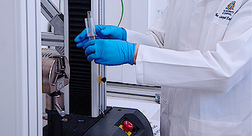
A. UNIAXIAL TESTING OF TISSUE
Uniaxial testing is suitable for tissues that are subjected to this form of loading in vivo, such as the ligaments, tendons and muscles. Specimens are extracted from fresh tissue and connected to a set of mechanical clamps that can control the length of the tested strip. Specimens are mounted within a small tank filled with a preservation medium to protect against dehydration during the tests.

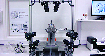
B. Biaxial testing of tissue
A biaxial loading instrument has been designed and is being manufactured by the Biomechanical Engineering Group. The instrument will enable the testing of square or rectangular tissue specimens (with a range of aspect ratios up to 2:1) and the application of tension or compression loads in two orthogonal and independent directions. Once ready, the rig will be used to test tissue specimens under biaxial loadings, hence making the loading regime acting on the tissue more representative of physiologic conditions than uniaxial testing.
C. Inflation testing of ocular tissue
Inflation testing has been used for more than 40 years. The test is considered superior to uniaxial and biaxial testing methods as it simulates better the in vivo conditions. Our inflation test rig has been designed and built in house. The rig enables testing of whole ocular globes under an internal pressure simulating the intraocular pressure (IOP). The rig provides tight control of hydration, temperature and pressure application rate, is computer-controlled and uses non-contact behaviour monitoring techniques. The systems enable a high level of user control and high reliability in results. The material properties obtained from this rig have been embodied in our predictive numerical simulations of ocular behaviour and enabled a close match with experimental response to mechanical actions.
D. Results
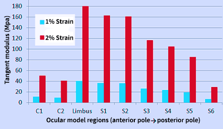
d.i. Regional variation of ocular biomechanical stiffness
Testing intact eye globes under inflation conditions enabled the determination of the regional variation of mechanical properties across the ocular surface. A first step in this work considered the eye to be divided into segments.
The stress-strain behaviour obtained for each segment enabled the determination of the tissue’s tangent modulus at specific strains or stresses.
In both human and porcine eyes a consistent trend has been observed with the central cornea exhibiting stiffer behaviour compared to the peripheral cornea, and the limbus showing a high stiffness, reducing gradually towards the posterior pole.
D.ii. Regional variation of ocular behaviour based on microstructure data
A study was conducted to numerically represent the regional and directional variation in mechanical stiffness (using X-ray scattering data describing the microstructure) across the human cornea and sclera. The data was analysed to determine the regional variation of stiffness and the anisotropy with a high level of control down to individual elements in a finite element model (FEM) of the eye. Bespoke software was developed to process the wide angle X-ray scattering (WAXS) data as shown in figure, and the resulting microstructure data was read automatically by our finite element analysis software to control the material properties at each model element.
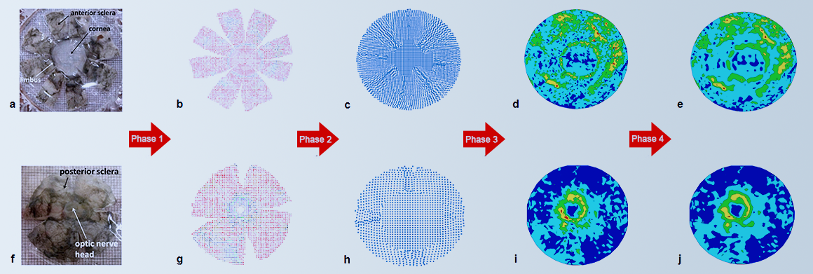
The complete process of attaining collagen fibrils density and distribution across the ocular surface of the human eye. The Anterior cup (top) is represented by figures (a) through (e), while the Posterior cup (bottom) is represented by figures (f) through (i). The blocks below explain each process briefly.
Prior to the WAXS method, the intact human specimen undergoes dissection according to the flattened layout presented in figures (a) and (f). WAXS is then used to determine fibril density and angular distribution at points with a 0.5mm grid spacing.
Using bespoke software, all the data points were analyzed and the maps were expanded to eliminate all gaps artificially created during dissection. The results are shown in figures (c) and (h).
The data collected at discrete points were separated into 6 maps representing the fibril density, isotropic distribution, two main angles of anisotropic distribution and the associated fibril densities.
Each of the 6 maps was fitted to Zernike polynomials (with a high order of 60) to remove global noise in data and sudden changes in data values that could cause instability in numerical modelling.
d.ii.1. Viscoelasticity of ligament tissue
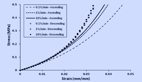
In this study three strain rates (0.1, 1 and 10%/min) were applied, to correspond to a gradual increase and decrease in the motion of the tibia relative to the femur. These tests were performed in ascending and descending order, When the motion decreased from fast to slow, the CCL behaviour was stiffer than when the motion increased from slow to fast (Figure 1). This might be one of the explanations of why warm-up exercises are important to reduce risks of knee injury in athletes.
comparison graph to show the difference in stress-strain behaviour of CCL during ascending and descending tests.
d.iii. Age-related stiffening of ocular tissue
A study was conducted to numerically represent the regional and directional variation in mechanical stiffness (using X-ray scattering data describing the microstructure) across the human cornea and sclera. The data was analysed to determine the regional variation of stiffness and the anisotropy with a high level of control down to individual elements in a finite element model (FEM) of the eye. Bespoke software was developed to process the wide angle X-ray scattering (WAXS) data as shown in figure, and the resulting microstructure data was read automatically by our finite element analysis software to control the material properties at each model element.
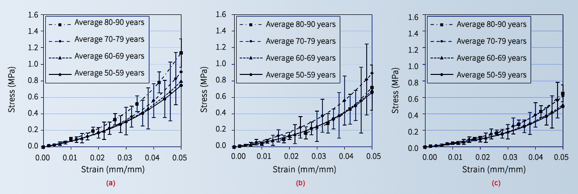
Comparison of average stress-strain behaviour within the four age groups for (a) anterior specimens, (b) equatorial specimens, and (c) posterior specimens. Error bars depict the standard deviation of stress values.
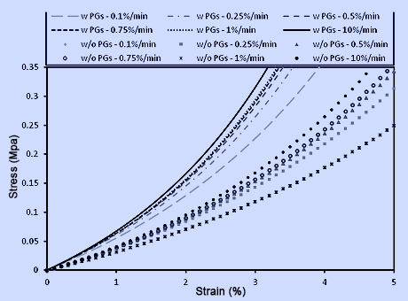
d.iv. Effect of proteoglycan content on behaviour of knee joints
Proteoglycans (PGs) are minor extracellular matrix proteins. The contribution of PGs to the viscoelasticity of ligaments such as the cranial cruciate ligament (CCL) in dog, or anterior cruciate ligament (ACL) in man has not been determined to date. In this study we are aiming to address the hypothesis that PGs play a vital role in biomechanical behaviour of ligaments such as viscoelasticity of the CCL.
In our study we have found that ligaments with depleted PGs had a reduced stiffness compared to ligaments with normal PG content.
Comparison graph to show the difference in stress-strain behaviour of CCL with and without proteoglycans..
OUR MAIN RESEARCH AREAS:
The Group carried out extensive tests to determine the mechanical properties of soft tissues.
more infoOur simulations represent closely the mechanical properties of materials and the organ’s topography.
more infoWe are involved in the developed of a number of medical devices to improve clinical practice.
more infoMathematical algorithms have been developed to improve customisation of surgical procedures.
more info